Compute Your FIB-4 Score Before Trusting a Normal ALT
Compute your FIB-4 score from bloodwork you already have, see why normal ALT misses fatty liver, and run a quantified diet protocol to reverse early MASLD.
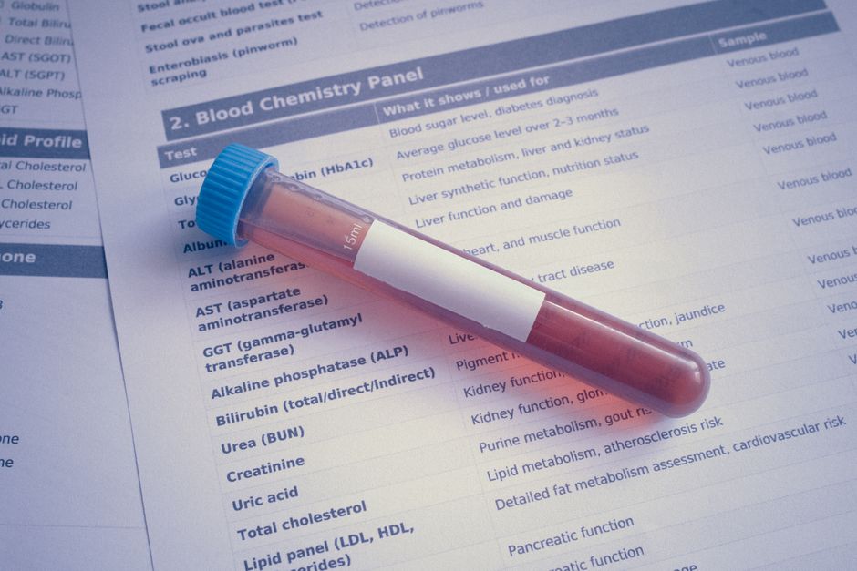
Your liver is the first organ to collect metabolic damage and the most forgiving one if you catch it early, yet the panel you already order twice a year quietly passes on it. A CMP reads your ALT, sees 22 U/L, and files the liver under "fine." That is weak evidence, and hepatology has known it for years. The fix costs nothing: a FIB-4 score computed from the age, AST, ALT, and platelet count sitting in the CMP and CBC you already order. Below you will find three worked calculations, the cut-offs that actually matter, explicit rules for when to escalate to elastography, and a reversal protocol written as doses. Grams of fructose, units of alcohol, percent of body weight, protein per kilogram, rather than another list of foods to avoid.
Stay in the loop.
Get the latest posts and exclusive content delivered to your inbox.
Join 37 readers. No spam. Unsubscribe in one click, anytime.
Why the Liver Shows Metabolic Damage First
MASLD, metabolic dysfunction-associated steatotic liver disease, affects somewhere between a quarter and a third of adults worldwide by most estimates, and the overwhelming majority of them feel nothing. Two properties make it a perfect target for a tracker. First, exposure: everything absorbed through the gut drains into the liver through the portal vein, so hepatic tissue sees dietary fructose and chylomicron remnants before any other organ does. Under insulin resistance, visceral fat also exports free fatty acids to the liver around the clock. The organ is downstream of your worst metabolic decisions and has nowhere to hide.
Second, asymmetry. Steatosis itself is close to fully reversible, and even early-stage fibrosis can regress, which is rare in human pathology. Catch the problem as fat and you can delete it; catch it as cirrhosis and you are negotiating for transplant time. The variable that separates those futures is fibrosis stage, and fibrosis stage is exactly what FIB-4 estimates.
Compute Your FIB-4 Score From a Panel You Already Have
FIB-4 = (Age × AST) ÷ (Platelets × √ALT)
- Age in years
- AST and ALT in U/L
- Platelets in 10⁹/L with SI units or ×10³/µL with US convention; the numeric value is identical, so read it straight off the CBC
- Thirty seconds and a calculator with a square-root key
The score was derived in 2006 to stage fibrosis without biopsy and has since been validated in MASLD populations; the AASLD 2023 practice guidance recommends it as the first-line noninvasive fibrosis risk test for people with metabolic risk factors, which describes most of a self-tracking readership. Three realistic panels:
| Panel | Age | AST | ALT | Platelets | Calculation | FIB-4 |
|---|---|---|---|---|---|---|
| A, lean tracker, 42M | 42 | 24 | 28 | 250 | (42 × 24) ÷ (250 × √28) = 1008 ÷ 1322.9 | 0.76 |
| B, 55F, mild metabolic flags | 55 | 32 | 38 | 210 | (55 × 32) ÷ (210 × √38) = 1760 ÷ 1294.5 | 1.36 |
| C, 68M, longtime lifter | 68 | 30 | 26 | 195 | (68 × 30) ÷ (195 × √26) = 2040 ÷ 994.3 | 2.05 |
Before the cut-offs, learn how the machine thinks, because the variable sensitivities are the whole story of liver blood panel interpretation:
- Platelets dominate. They enter the denominator linearly, because fibrosis raises portal pressure, which swells the spleen, which sequesters platelets. Drop Panel A's platelets from 250 to 190 with a medication effect or an unrelated thrombocytopenia and the score climbs from 0.76 to 1.00 with the liver untouched.
- A rising ALT lowers the score. Double Panel A's ALT to 56 and FIB-4 falls from 0.76 to 0.54, because ALT sits under a square root in the denominator. FIB-4 is an architecture score, not an injury score; a hot enzyme flare can partially mask the fibrosis signal.
- Age inflates mechanically. Calendar time is a numerator term. The same pristine liver scores about 75 percent higher at 70 than at 40, which is why the cut-offs change with age.
- AST is not liver-specific. Hard eccentric training leaks muscle AST into serum for days. Do not draw within several days of a brutal session, or you will agonize over a training artifact.
FIB-4 Risk Tiers, Cut-offs, and Honest Limitations
| FIB-4 value | Adults under ~65 | Adults over ~65 |
|---|---|---|
| Below 1.30 (below 2.00 if 65+) | Low risk | Low risk |
| 1.30 to 2.67 (2.00 to 2.67 if 65+) | Indeterminate | Indeterminate |
| Above 2.67 | High risk | High risk |
A low FIB-4 is real reassurance, with negative predictive values for advanced fibrosis commonly reported near 90% in validation cohorts. An indeterminate value is not reassurance; guidelines route it to second-line testing. Expect to land there, because a large share of screened patients fall in the middle band, which is a limitation of the tool, not a verdict about you. A high value demands action, detailed two sections down.
The 2.00 cut-off for older adults exists because age inflates the numerator; age-adjusted FIB-4 thresholds trade some sensitivity for far fewer false alarms. Panel C illustrates the flip side: 2.05 at age 68 is indeterminate under both rules, but it clears the age-adjusted 2.00 floor by a hair instead of sitting mid-band. The cut-off earns its keep a tier lower, where an older adult at 1.70 is low risk under the age-adjusted rule and indeterminate under the younger one. Other failure modes worth knowing: non-hepatic thrombocytopenia (immune thrombocytopenia, medications, B12 or folate deficiency), acute inflammation elevating AST, muscle-derived AST, and some evidence the score discriminates less well in type 2 diabetes. Diabetes is a reason to lower your escalation threshold, not to raise your reassurance.
Why a Normal ALT Does Not Clear You
ALT is a leak marker. Enzymes escape into serum when hepatocytes rupture, so ALT measures cell death happening now, not the fat accumulating quietly or the collagen being laid down around sinusoids. A steatotic liver can be badly dysfunctional while its cells die slowly enough to keep ALT inside the range.
Two compounding problems make the standard flag system worse. The reference range itself is contaminated: the familiar upper limits near 40 U/L for men were set from populations that included undiagnosed fatty liver and viral hepatitis, and many hepatologists have argued for decades that honest limits sit closer to 30 U/L for men and in the low twenties for women. The epidemiology agrees with the skepticism. Cohort data on normal ALT consistently show that a large share of people with MASLD, including a meaningful minority of those with advanced fibrosis, hold normal aminotransferases. So, can you have fatty liver with normal ALT? Yes, and the normal-ALT subgroup is arguably where tracking helps most, because nobody is going to accidentally investigate you.
The practical move for panel readers is to treat ALT as a trend, not a pass/fail. A stable 19 and a drifted 38 both print without flags. They are not the same liver. Direction inside the reference range carries information your lab report throws away.
When to Escalate to Elastography
Noninvasive liver fibrosis tests form a ladder, and elastography is the second rung. Vibration-controlled transient elastography (FibroScan) sends a shear wave through the liver, reports stiffness in kPa that maps to fibrosis stage, and adds a controlled attenuation parameter (CAP) that grades steatosis. Fifteen minutes, no needles.
Escalate when any of these hold:
- FIB-4 at or above 1.30 (at or above 2.00 if over 65) on a confirmed draw. Repeat the CMP and CBC weeks later, outside any hard training block. If it holds, request VCTE or an enhanced liver fibrosis (ELF) serum panel.
- FIB-4 above 2.67. Repeat once to exclude artifact, then go straight to a hepatology referral rather than imaging alone.
- Metabolic red flags at any score. Type 2 diabetes, a metabolic-syndrome triglyceride and HDL picture, a platelet count trending down across years, or a family history of cryptogenic cirrhosis all justify imaging at a lower bar.
Do not escalate when the score sits low, enzymes are quiet, and the metabolic cluster is absent. That is retest-annually territory, and scanning it anyway mostly buys anxiety. If you do request it, a workable script:
"I'd like vibration-controlled transient elastography to assess liver fibrosis risk. My FIB-4 is X on two draws; if the scanner is unavailable, an enhanced liver fibrosis panel is an acceptable alternative."
Two honest caveats. Stiffness readings degrade in severe obesity, where the XL probe helps, and acute inflammation or cardiac congestion can inflate kPa independent of fibrosis. MRI-PDFF remains the most sensitive liver-fat quantification and is increasingly available cash-pay if you want research-grade steatosis numbers.
The Quantified Reversal Protocol

Assume elastography or a strong risk picture shows early disease: steatosis, possibly stage 1 fibrosis. Fatty liver reversal is not a mystery; it has a dose-response, and almost all of it belongs to one lever. Weight loss is the only intervention with trial-backed dose-response evidence for fibrosis itself. Everything else is either a gate (alcohol, sugar-sweetened beverages) or a support structure (protein, the Mediterranean pattern). Ranked by evidence strength and effect per unit of effort, with the metric each lever should move:
| Rank | Lever | What the evidence supports | Metric it moves | Clock |
|---|---|---|---|---|
| 1 | Weight loss, 7 to 10% of body weight | Dose-response for steatosis, NASH resolution, fibrosis | CAP first, kPa later | Fat in weeks, fibrosis in months |
| 2 | Zero sugar-sweetened beverages, added sugar under ~25 g/day | Liver fat falls within weeks of cutting liquid fructose | CAP | Weeks |
| 3 | Alcohol at zero for the window | No established safe threshold; protects the gains from levers 1 and 2 | Progression risk | The whole window |
| 4 | Protein at 1.2 to 1.6 g/kg/day | Lean-mass retention and deficit adherence | Waist, weight trend | Weeks to months |
| 5 | Coffee at 2 to 3 cups/day | Lower fibrosis risk, association only | None you can bank on | Background |
Weight: the only lever that touches fibrosis. Around 5 percent of body weight is where steatosis measurably improves; randomized weight-loss trial evidence associates roughly 7 percent with NASH resolution and about 10 percent with fibrosis improvement. For a 90 kg reader that is 6.3 to 9 kg. Pace it at 0.5 to 1 percent of body weight per week, which lands the full loss in roughly two to five months while sparing lean mass. Faster deficits mostly burn the muscle you will want back.
Fructose: a gate, not a therapy. Fructose reaches the liver first, bypasses the phosphofructokinase checkpoint that throttles glucose metabolism, and feeds de novo lipogenesis directly; controlled fructose-restriction trials show liver fat falling within weeks of cutting it, particularly from sugar-sweetened beverages. Operational caps: zero sugar-sweetened beverages during the reversal window, and added sugar under roughly 25 g per day, the neighborhood of major guideline limits. Whole fruit is not the demonstrated problem; liquid and added sugar are. Holding the gate stops a driver; it does not substitute for the deficit.
Alcohol: zero for the window. No safe intake threshold for MASLD progression has been established, and guidelines advise minimal to no intake while reversing steatosis. A defined 6 to 12 month abstinence window is cleaner than a "cut back" that quietly drifts.
Protein: support structure. A protein-forward, lower-refined-carbohydrate structure defends lean mass during the deficit and makes the deficit tolerable, and randomized Mediterranean-diet evidence supports the broader pattern for liver-fat reduction specifically. Anchor each meal with protein, take carbohydrate from whole sources, and let fat come mostly from olive oil and nuts.
Coffee: an association, ranked last for exactly that reason. Observational and meta-analytic data link 2 to 3 cups daily with lower fibrosis risk. A favorable correlation with a cheap downside profile, never a proven cause.
Read the clocks, not just the doses: the CAP side of the scan responds in weeks, which is what the fructose gate and the first kilograms of weight loss should move; the kPa side shifts over months and only meaningfully after the full 7 to 10 percent lands. The retest cadence below is built on that split.
Retest Cadence and Tracking Reversal
Different tissues move at different speeds, so match the metric to the clock. Steatosis shifts in weeks to months; fibrosis takes months to years; and FIB-4, validated as a one-time gate, makes a noisy serial monitor. Do not over-read a drift from 1.36 to 1.24.
- Every 3 months: CMP plus CBC (ALT, platelets) and recompute the score. The platelet trend is quieter and often more meaningful than ALT, since rising platelets can reflect easing portal pressure.
- Monthly: waist circumference at the navel, a proxy for the visceral depot driving free-fatty-acid flux to the liver.
- At 6 to 12 months: repeat VCTE with CAP. Success is a full steatosis-grade improvement; if you bought a baseline MRI-PDFF, judge the relative liver-fat drop rather than the absolute grade.
- Annually if low risk: FIB-4 plus ALT is cheap permanent insurance.
Prove reversal on paper. A number you never re-measured is a hope, not a result.
The HackedSelf Liver Protocol Card
| Phase | Action | Threshold or number |
|---|---|---|
| 1. Assess | Compute FIB-4 from your last CMP | (Age × AST) ÷ (Platelets × √ALT) |
| 2. Interpret | Tier the score | <1.30 low (<2.00 over 65); 1.30 to 2.67 indeterminate; >2.67 high |
| 3. Escalate | VCTE or ELF at ≥1.30 (≥2.00 if 65+); hepatology above 2.67 | Confirm on a repeat draw first |
| 4. Dose | 7 to 10% body-weight loss; zero SSBs; added sugar under ~25 g/day; alcohol zero for the window; protein 1.2 to 1.6 g/kg; coffee 2 to 3 cups | 0.5 to 1% body weight per week |
| 5. Retest | CMP and FIB-4 every 3 months; VCTE with CAP at 6 to 12 months; waist monthly | Steatosis-grade drop equals success |
Few organs forgive the way the liver does, and almost none forgive as completely at the fat stage. The four numbers that tell you which stage you are in are already in your inbox. Compute the score before you trust the ALT.
Stay in the loop.
Get the latest posts and exclusive content delivered to your inbox.
Join 37 readers. No spam. Unsubscribe in one click, anytime.
About the author
Jordan Reyes
Registered Dietitian
Jordan ditched diet dogma for metabolic health, running continuous glucose monitors and food journals to see what actually moves the needle. He writes nutrition and supplement protocols grounded in evidence, not influencer trends.
Related Posts

Artificial Sweeteners Gut Bacteria Risks Across 39 Types
Artificial sweeteners gut bacteria research shows 39 sweeteners act differently when combined with caffeine and medications. Find your safest options.

Low-Fat Vegan Diet Type 2 Diabetes Trial, an Omnivore's Audit
A mechanism audit of the low-fat vegan diet type 2 diabetes trial, plus the fat, fiber, and cooking swaps omnivores can steal for better blood sugar.
4 to 6 Whole Grain Servings Per Day, Dosed in Grams
Whole grain servings per day, translated into grams. Turn the 4 to 6 servings from 87 trials into a protein-first heart health protocol you can weigh.